
Sensing Knife

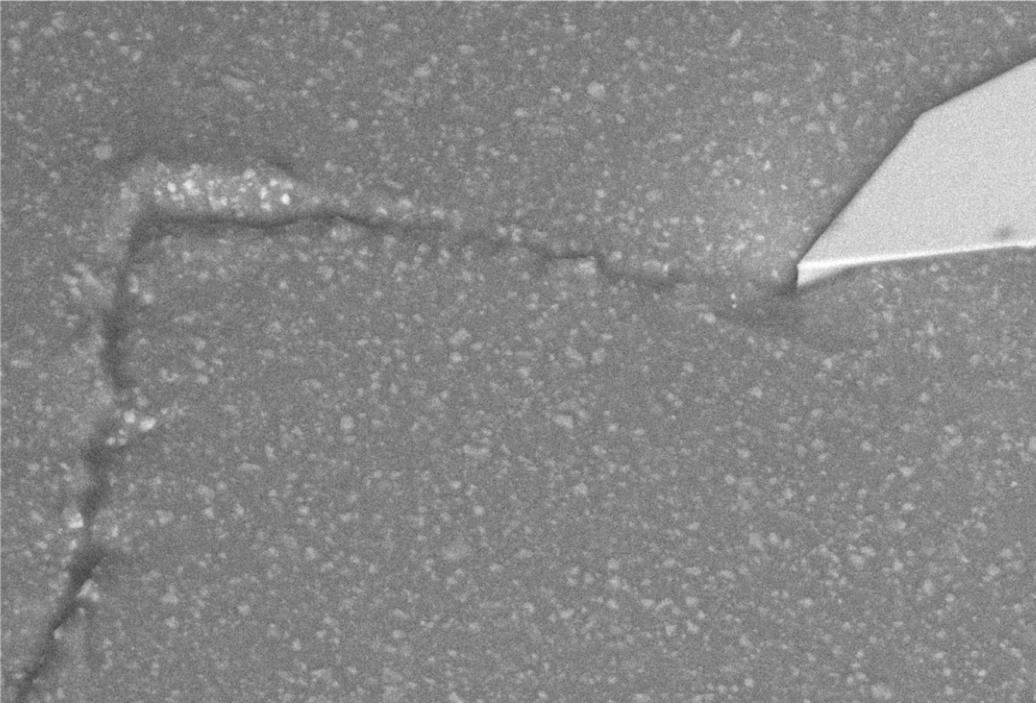
Scanning electron microscope image of the scanning probe microscope Silicon nitride sharp probe. At 99300x magnification. An apex radius of just 7 nm is typical. Copyright: Mohammad Fardin Gholami and Johannes Müller
Achieving a minimal level of lesion is often essential when cutting and manipulating materials. For this, we foresee an immense advance with the invention of cutting tools able to detect and distinguish materials based on their properties at the relevant scales. The Sensing Knife project aims at utilising the physical working principles of Scanning Probe Microscopy (SPM)(*1,2) will be used to examine active matter (*3-5) in its delicate, dynamic and functional, e.g. brain matter, or fundamental, e.g. molecular scale. Within this framework, our objective is to achieve precise recognition and manipulation of tissues & materials at scales of a few hundred of micrometres (smaller than the precision of surgeons' hands) and to explore matter and its functional properties simplified as healthy or unhealthy, e.g. tumour cells or wanted and unwanted.
Sensing Knife project has been focused on 1) Collaborative work with virtual environment artists for creating VR interactives of virtual sensing knife for future cutting planning processes, 2) creation and optimising prototypes for cutting by integrating both sensing and simultaneous cutting based on scanning probe microscopy and spectroscopy principles and 3) advancing prototypical and spectroscopic methods on sensing and manipulating patients tumour resections (explant tissues). We will further develop our new sensing knife prototype and data analysis and visualisation methods to experimentally navigate in-between areas of active matter examples using physical principles acting on matter and probes.
Based on those sensing probes, we imagine developing methods and tools to intervene at molecular and cellular scales. As a result, we envision the reduction of energy costs, time and error rates, as well as dramatically improved recovery processes in micro and Nano surgical environments for specialised treatments to specific conditions. For this purpose, 1) we will use experimental data gathered by SPM force spectroscopy and indentation experiments to describe the physical variations of living healthy vs. cancerous glial cells and interfaces between them. 2) The data will be further compared to the tissue scale of active matter e.g., brain matter and tumour explants and its healthy tissue interface. 3) A »Sensing Knife« prototype will be further modified to work within the required ranges of sensitivity and detection suitable for real-world biological matter. In parallel, we will further establish and improve the SK prototype by publishing the instrumentation and methodology and testing its functionality and impact within a robotic positioning system as a direct collaboration with the project Robotic-assisted Surgery.
Literature
1) Proksch, R. et al. Practical loss tangent imaging with amplitude-modulated atomic force microscopy. J. Appl. Phys. 119, 134901 (2016).
2) Herruzo, E. T., Perrino, A. P. & Garcia, R. Fast nanomechanical spectroscopy of soft matter. Nat. Commun. 5, 3126 (2014).
3) Gholami, M. F., Guiducci, L., Jany, S. & Razghandi, K. Rethinking Active Matter: Current Developments in Active Materials. in Active Materials (eds. Fratzl, P., Friedman, M., Krauthausen, K. & Schäffner, W.) 191–222 (De Gruyter, 2021). doi:10.1515/9783110562064-011
4) Efremov, Y. M., Wang, W.-H., Hardy, S. D., Geahlen, R. L. & Raman, A. Measuring nanoscale viscoelastic parameters of cells directly from AFM force-displacement curves. Sci. Rep. 7, 1541 (2017).
5) Mohammadifar, E. Gholami, M. F. et al. Graphene‐Assisted Synthesis of 2D Polyglycerols as Innovative Platforms for Multivalent Virus Interactions. Adv. Funct. Mater. 31, 2009003 (2021).